POCUS Tips › Lung
Normal Lung — A-ProfileA-LINE
Learn the baseline first: sliding pleura plus A-lines means air where air belongs, and it is what every pathology is compared to.
Open the interactive view with the loop and quiz →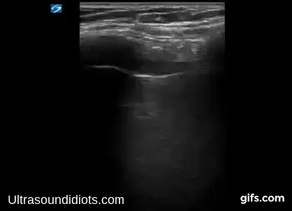
Where to put the probe
- Window
- Anterior chest, mid-clavicular line, probe across two ribs
- Marker
- Patient’s HEAD (12 o’clock)
- probe
- linear
- preset
- lung
- depth
- 4–6 cm
Get the view
- Hold the probe longitudinally, perpendicular to the ribs: two rib shadows with the pleural line between them is the bat sign.
- Center the pleural line and reduce depth until it occupies the top third of the screen.
- Watch through one full breath for shimmer along the pleura — that is lung sliding.
- Then work the zones in order: anterior upper, anterior lower, lateral, and posterolateral at the base.
What you are looking at
- Soft tissue and rib
- Layered muscle and fascia in the near field; ribs cast clean posterior shadows — the wings of the bat.
- Pleural line
- The bright horizontal line where parietal meets visceral pleura — the reference for everything else on the screen.
- Lung sliding
- Shimmering to-and-fro motion along that line with respiration. Present means no pneumothorax at that spot.
- A-lines
- Equidistant horizontal repeats of the pleural line — normal reverberation through air.
- Seashore sign
- The M-mode signature of sliding: grainy sand under a smooth sky. Side-by-side on the pneumothorax page.
What normal looks like
| Sign | Normal | Notes |
|---|---|---|
| Bat signlongitudinal, across two ribs | present | if it is missing you are lying along a rib — rotate 90° |
| A-linesspacing equals skin-to-pleura distance | repeating | A-lines plus sliding rules out pneumothorax and interstitial fluid at that spot |
| B-linescount per intercostal space | ≤ 2 | ≥ 3 in one space is a positive zone, especially anteriorly |
| M-modesingle line through the pleura | seashore | barcode pattern means no sliding |
Don’t be fooled — pitfalls
- Subcutaneous emphysema abolishes everythingAir in the soft tissue blocks the beam and mimics pneumothorax. If the near field is a mess of bright noise, do not interpret the pleura.
- Lung pulse is not slidingCardiac motion transmitted to the pleura shows movement without true sliding — think atelectasis or mainstem intubation, not pneumothorax.
- Scanning below the diaphragmAt the bases it is easy to image liver or spleen and call it consolidated lung. Find the diaphragm and stay above it.
- Too much depthDeep settings shrink the pleural line to a few pixels and hide the detail you are looking for.
Bad image? Fix it
- No bat signYou are parallel to a rib. Rotate 90° so the footprint crosses two ribs.
- Cannot tell if it is slidingFreeze the debate with M-mode: seashore is sliding, barcode is not.
- Noisy near fieldReduce overall gain, drop near-field TGC, and prefer the linear probe for pleural detail.
- Need to see effusion or consolidation tooSwitch to curvilinear or phased array at 10–12 cm — the linear probe will not reach.
So what — what the finding changes
- A-profile with sliding, in a hypoxic patientCardiogenic pulmonary edema becomes unlikely and pneumothorax is excluded at the spots you scanned — pivot to COPD, asthma, or PE, and check the posterolateral bases: an A-profile with a posterior consolidation or effusion (PLAPS) points to pneumonia.
- A-profile without slidingA-lines alone do not rule out pneumothorax — they persist in PTX too. Absent sliding is the finding that matters; go hunt for a lung point.
- A-profile in undifferentiated shockLungs are not the answer. Move to the heart, IVC, abdomen and aorta.
- Bilateral A-lines after intubationConfirms aerated lung, but check for equal sliding — absent sliding on the left suggests mainstem placement.
Related views
Keep going
Educational use only. Not for diagnosis or treatment decisions — confirm findings with formal imaging and your own judgment. By Yonathan Daniel, MD · Pathways · Quiz on this view · About · Last reviewed 2026-10-04.