POCUS Tips › Abdomen
Abdominal AortaAORTA
Rule an abdominal aortic aneurysm in or out at the bedside: the whole aorta from diaphragm to bifurcation, measured outer wall to outer wall.
Open the interactive view with the loop and quiz →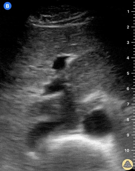
Where to put the probe
- Window
- Midline epigastrium, then sweep caudally to the umbilicus
- Marker
- Patient’s RIGHT (transverse, ~9 o’clock); rotate to the HEAD for the long axis
- probe
- curvilinear
- preset
- abdominal
- depth
- 15–20 cm
Get the view
- Transverse just below the xiphoid, firm steady pressure. Find the vertebral body’s bright arc with its shadow; the aorta sits just anterior and to the patient’s left of it, the IVC to the right.
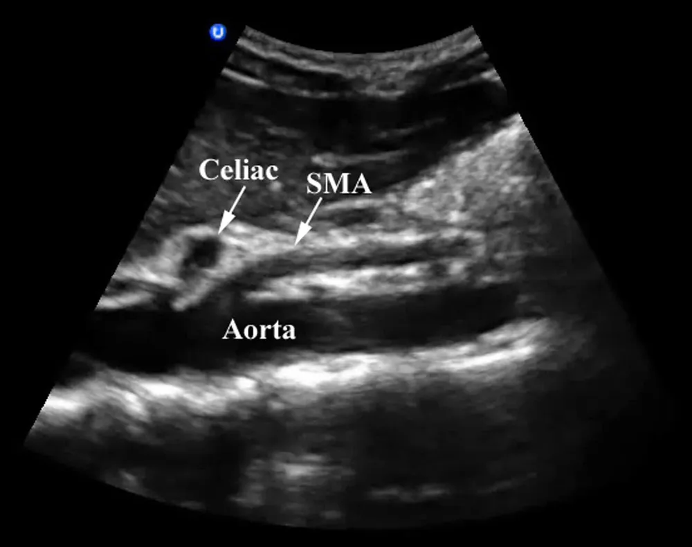
- Identify the proximal aorta by its branches: the celiac trunk, then the SMA running parallel below it. Confirm it is the aorta — thick-walled, round, pulsatile, non-compressible.
- Sweep caudally in transverse, keeping the aorta centred, past the renal arteries to the bifurcation at about the umbilicus. Measure the maximum AP diameter outer wall to outer wall at the widest point.
- Rotate to the long axis (marker to the head) and confirm the diameter and the wall, then follow the common iliacs for a short distance.
What you are looking at
- Aorta
- Round, thick bright wall, pulsatile, just anterior and to the patient’s left of the vertebral body. Calipers go outer wall to outer wall.
- IVC
- To the patient’s right (screen left): oval, thin-walled, changes calibre with breathing and flattens with probe pressure. Never measure this one as the aorta.
- Vertebral body
- The bright arc with a shadow behind it — your landmark. If you cannot see it, you are not deep enough or gas is in the way.
- Liver (acoustic window)
- The left lobe fills the near field and carries the beam to the vessels; the black branches inside it are hepatic and portal veins, not the IVC.
- Celiac trunk
- First anterior branch, just below the diaphragm — the seagull sign in transverse. Shown on the long-axis still below.
- Superior mesenteric artery
- Just below the celiac; runs parallel to the aorta in the long axis. The renal arteries arise just below it.
- Bifurcation
- At roughly the umbilicus. Most AAAs are infrarenal, so the segment just above this is the one you cannot skip — see the sweep below.
On the screen
- 1Aorta (coronal)
- 2IVC
- 3Celiac trunk
- 4Superior mesenteric artery
- 5Bifurcation
- 6Aorta (transverse)
- 7Vertebral body
- 8IVC (transverse)
Numbers that matter
| Measure | Normal | Call it abnormal when |
|---|---|---|
| Aortic AP diameterouter wall to outer wall, transverse, widest point | < 3.0 cm | ≥ 3.0 cm is an aneurysm; ≥ 5.0–5.5 cm is the usual repair range |
| Symptomatic aneurysmany size + pain, syncope or hypotension | — | treat as rupture until proven otherwise — the size does not reassure you |
| Common iliac diametertransverse, below the bifurcation | < 1.5 cm | ≥ 1.5 cm is aneurysmal |
| Segments visualisedproximal, mid, distal, bifurcation | all four | any segment not seen makes the study inadequate — say so and get CT |
Don’t be fooled — pitfalls
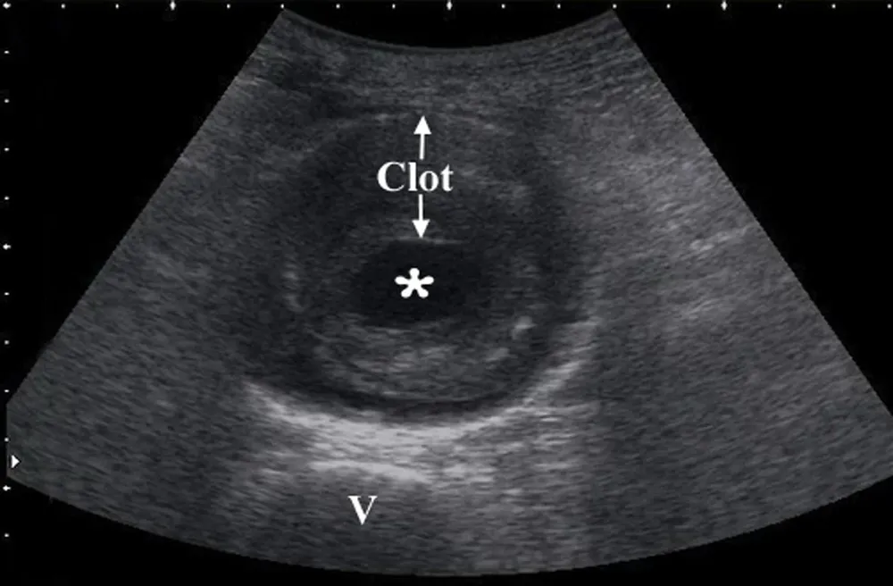
- Measuring the lumen inside thrombusMural thrombus is echogenic and can look like a wall. Calipers go from outer wall to outer wall or you will under-call a large aneurysm.
- IVC mistaken for the aortaThe IVC is on the patient’s right, oval, thin-walled, flattens with probe pressure and changes with breathing. Trace the vessel to a branch (celiac, SMA) or the heart before you measure.
- Oblique cuts overestimateA tangential transverse plane makes a round vessel an oval. Measure the short axis of that oval, and confirm in the long axis.
- Rupture is retroperitonealBlood from a ruptured AAA usually tracks into the retroperitoneum where ultrasound cannot see it. A negative FAST does not exclude rupture.
- A tortuous aorta drops out of planeOlder aortas bow to the left. Follow the vessel with small rotations rather than sliding straight down the midline.
Bad image? Fix it
- Bowel gas over the aortaGraded, sustained pressure to push gas aside; wait a few breaths; or come in from the right flank using the liver and IVC as a window.
- Everything is too deep and darkLower the frequency, increase depth until the vertebral body appears, then set the focal zone at the aorta.
- Lose the vessel in the long axisReturn to transverse, centre the aorta, then rotate slowly keeping the centre fixed.
- Large or distended abdomenKnees bent to relax the wall, left lateral decubitus, and the flank approach.
So what — what the finding changes
- AAA + hypotension or painRuptured until proven otherwise: vascular surgery and blood now. An unstable patient goes to theatre, not to the CT scanner.
- Symptomatic aneurysm, stableCT angiography and urgent vascular review in the same visit.
- Incidental aneurysm ≥ 3 cmDocument the size and refer for surveillance; counsel the patient on rupture symptoms.
- Cannot see the whole aortaAn inadequate study is not a negative study. Document the limitation and image another way if the question matters.
Keep going
Educational use only. Not for diagnosis or treatment decisions — confirm findings with formal imaging and your own judgment. By Yonathan Daniel, MD · Pathways · Quiz on this view · About · Last reviewed 2026-10-04.